Introduction to Murine Hepatitis Virus (MHV)
Murine Hepatitis Virus (MHV) is a coronavirus that primarily affects laboratory mice and is an essential tool in virology and immunology research. This virus is often used as a model organism to study human coronavirus infections, including those caused by viruses such as SARS-CoV-2. MHV plays a pivotal role in understanding viral replication, pathogenesis, and immune response. It also serves as a model for studying viral pathogenesis, immune evasion mechanisms, and antiviral therapies in the context of laboratory animal populations.
MHV is a positive-sense single-stranded RNA virus from the Coronaviridae family. It is genetically similar to human coronaviruses, making it a valuable surrogate for human coronavirus studies. In laboratory settings, it is crucial to maintain strict monitoring and diagnostic practices to ensure MHV does not contaminate research or impact the health of animal populations. Reliable detection and surveillance are essential, particularly when MHV has the potential to confound research results by causing inflammation, hepatic damage, or other pathological symptoms in infected animals.
Researchers use molecular diagnostic tools like Polymerase Chain Reaction (PCR) to detect MHV. Accurate detection is essential for preventing outbreaks in animal facilities and ensuring contamination-free environments for experiments.
For comprehensive molecular and biological data on MHV, resources from the National Institutes of Health (NIH) (https://www.nih.gov) and PubMed (https://pubmed.ncbi.nlm.nih.gov) offer valuable scientific references.

Polymerase Chain Reaction (PCR) and Its Role in MHV Detection
Understanding PCR in Virology
Polymerase Chain Reaction (PCR) is an essential technique used in molecular biology for the amplification and detection of nucleic acid sequences. PCR amplifies small quantities of viral RNA or DNA, making it easier to detect specific sequences associated with pathogens such as Murine Hepatitis Virus. PCR testing is widely employed in diagnostic laboratories, especially when detecting viral infections like MHV.
The RT-PCR (Reverse Transcriptase PCR) process is particularly critical in detecting RNA viruses like MHV. RT-PCR converts RNA into complementary DNA (cDNA), which can then be amplified and detected. The amplification process is sensitive, allowing PCR assays to detect even minute quantities of viral RNA in clinical or research samples.
PCR assays can be divided into two categories:
-
Qualitative PCR — Aimed at detecting the presence or absence of a specific viral sequence. It confirms whether the virus is present in the sample but does not measure viral load.
-
Quantitative PCR (qPCR) — Involves measuring the amount of viral DNA/RNA present, providing a quantitative assessment of infection. This method is often used in viral load measurement in clinical diagnostics.
In MHV detection, qualitative PCR is more commonly used, focusing on identifying genetic markers of the virus without requiring quantitative measurements. The key advantage of qualitative PCR is its simplicity and ability to provide quick results that are vital for research protocols and animal health surveillance.
For additional understanding of PCR technology, you can explore NIH’s PCR resources (https://www.nih.gov) and NCBI’s molecular biology section (https://www.ncbi.nlm.nih.gov).
Critical Role of Quality Control in MHV PCR Detection
Ensuring the accuracy and reliability of PCR tests is critical in viral diagnostics. Quality control (QC) procedures must be followed meticulously to avoid errors that can lead to false positives or false negatives. False negatives can occur due to poor sample quality, inhibitors in the sample, or failure in the PCR amplification process. Conversely, false positives may result from contamination in the PCR setup, reagent kits, or laboratory environment.
To ensure the reliability of PCR results, various quality control measures must be implemented:
1. Use of Positive and Negative Controls
Positive and negative controls are essential for ensuring that the PCR assay functions correctly. A positive control consists of a known sample containing MHV RNA, ensuring that the PCR assay can successfully detect the target. A negative control involves testing a sample that should not contain MHV, ensuring that no contamination has occurred in the PCR reagents or setup.
2. Internal Controls
Internal controls are samples that help detect errors in the PCR process, such as poor extraction or PCR inhibition. These controls are crucial for confirming the success of the entire testing process and ensuring accurate results.
Laboratories are encouraged to implement rigorous protocols for control management as per FDA regulations and CDC guidelines, which specify how and when to use control materials (fda.gov).
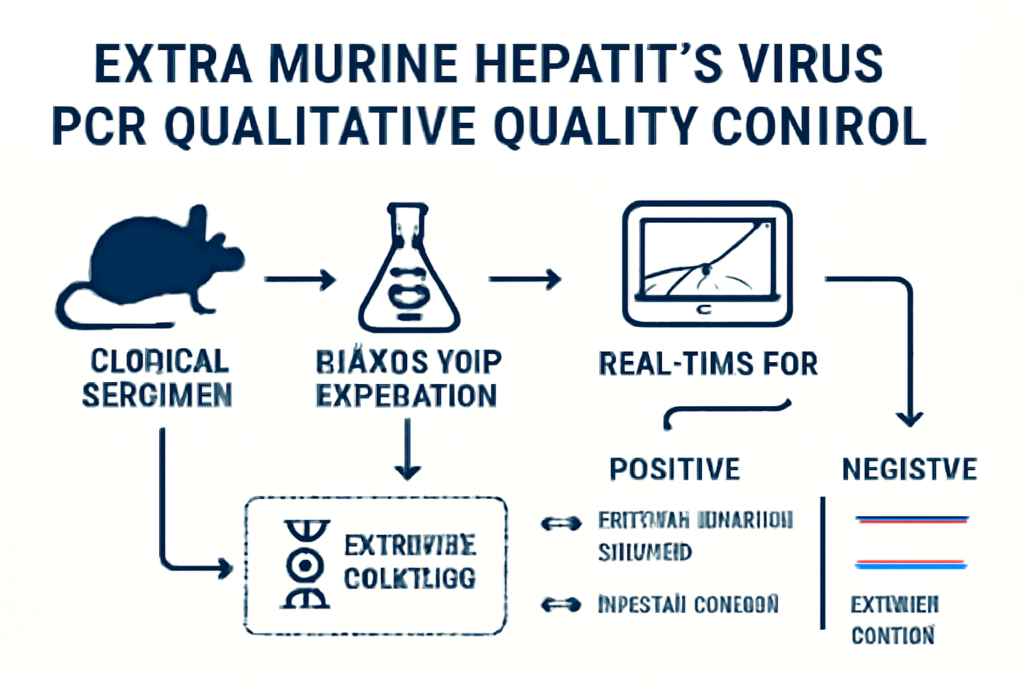
EXTRA MHV PCR Qualitative Quality Control Materials
EXTRA MHV PCR Qualitative Quality Control materials are specially designed reagents or synthetic samples that simulate real-life viral samples. These control materials are vital in validating PCR assay performance, ensuring that each PCR test is accurate, reproducible, and free from contamination. They include a variety of critical components such as:
-
Synthetic RNA Standards: These are used to simulate MHV RNA in test samples. They provide known quantities of the target RNA to ensure that the PCR system can detect the virus under controlled conditions.
-
Extracted Viral RNA: In some cases, laboratories use pre-extracted MHV RNA from infected murine samples to validate the PCR assay’s sensitivity and specificity.
-
Matrix Controls: These controls are used to validate the entire extraction and amplification system. They ensure that sample preparation, including RNA extraction, is functioning properly.
Benefits of Using Control Materials:
-
Validation: Ensures that the PCR assay works as intended and can accurately detect MHV.
-
Benchmarking: Provides a known standard against which experimental results can be compared.
-
Error Detection: Helps identify potential sources of error during the PCR process, such as contamination or inhibition.
The importance of control materials is outlined in FDA’s guidance for validating PCR assays in clinical laboratories (fda.gov).
Designing MHV-Specific PCR Assays
The success of PCR assays for MHV detection depends heavily on primer and probe design. These primers must target conserved regions of the MHV genome, ensuring that they amplify the viral RNA effectively without cross-reacting with host or other pathogens. Primers should be designed with the following considerations:
-
Specificity: Primers should be highly specific to MHV, ensuring they do not amplify any host or non-target viral sequences.
-
Amplicon Size: The amplified region should be small enough to be efficiently amplified by the PCR process while still being long enough to ensure detection accuracy.
-
Homology: Primers should be tested for homology across different MHV strains to ensure detection across the broadest range of MHV variants.
Bioinformatics tools such as BLAST (https://blast.ncbi.nlm.nih.gov) can be used to verify the specificity of primers and probes. The NIH Primer3 tool (https://primer3.ut.ee) can help design primers optimized for PCR.
Ensuring the Accuracy of PCR Results
PCR results must be accurately interpreted to ensure that the conclusions drawn from the data are correct. Several factors influence the interpretation of PCR results, including:
-
Amplification Efficiency: The PCR process must efficiently amplify the target sequence without degradation of the template.
-
Contamination: Laboratories must implement strict contamination control procedures to ensure the accuracy of results. This includes strict segregation of sample handling areas and frequent cleaning of PCR equipment.
-
Data Interpretation: Data generated from PCR assays must be carefully analyzed to ensure that signal thresholds are set appropriately to distinguish true positives from background noise.
The FDA’s Molecular Test Validation Guidelines provide a detailed framework for interpreting PCR results in diagnostic settings, including setting thresholds and validating assay sensitivity (fda.gov).
Advancements in MHV PCR Detection
As molecular techniques evolve, next-generation sequencing (NGS) is becoming an increasingly popular tool in MHV research and detection. NGS can provide a comprehensive view of viral diversity, enabling researchers to detect mutations in MHV strains that may influence the virus’s behavior or resistance to antiviral treatments.
Additionally, the use of real-time PCR has allowed for faster and more accurate monitoring of MHV viral loads, improving diagnostic timelines and the ability to track infection progression.
Conclusion: The Future of MHV PCR Detection
The future of MHV detection through PCR continues to advance with improvements in sensitivity, specificity, and speed. Through the use of high-quality control materials, optimized primer/probe design, and advanced diagnostic technologies, laboratories can improve the reliability and reproducibility of MHV PCR assays.
For research purposes, ongoing improvements in QC practices, the use of next-generation sequencing, and advancements in PCR technology will lead to better understanding of MHV pathogenesis and improved prevention and treatment strategies in research environments.
For further reading and detailed protocols, refer to trusted resources like the FDA, CDC, and NIH (cdc.gov, nih.gov).