Target overview: glucokinase (GCK)
Glucokinase catalyzes glucose + ATP → glucose-6-phosphate (G6P) + ADP. It is a distinct hexokinase IV isoform with unique kinetic properties (sigmoidal response to glucose and higher S₀.₅). See curated entries at NCBI Gene for GCK (gene ID, synonyms, transcripts) on NIH/NCBI: https://www.ncbi.nlm.nih.gov/gene/. For a controlled vocabulary on enzymes and metabolic pathways, check NLM MeSH terms: https://www.nlm.nih.gov/mesh/.
GCK reactions interface with carbohydrate metabolism; general biochemical pathway primers can be reviewed through NIH Bookshelf topic pages: https://www.ncbi.nlm.nih.gov/books/ and PubMed method overviews: https://pubmed.ncbi.nlm.nih.gov/.
Chemical identifiers (for reagents and standards):
-
Glucose (PubChem, NIH): https://pubchem.ncbi.nlm.nih.gov/compound/5793
-
ATP (PubChem): https://pubchem.ncbi.nlm.nih.gov/compound/5957
-
NADP+ (PubChem): https://pubchem.ncbi.nlm.nih.gov/compound/5884
-
NADPH (PubChem): https://pubchem.ncbi.nlm.nih.gov/compound/928
Assay principle (fluorometric, coupled detection)
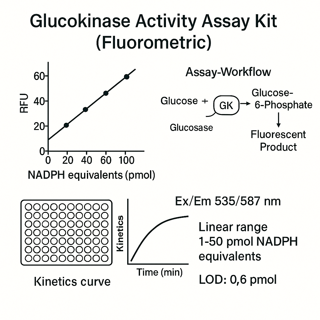
The Glucokinase Activity Assay Kit (Fluorometric) typically measures GCK activity by coupling G6P production to a fluorescent readout. Two common coupling strategies are used in research:
-
Dehydrogenase coupling: G6P is oxidized by G6PDH in the presence of NADP+, generating NADPH, which is intrinsically fluorescent (commonly measured near Ex ~ 340 nm / Em ~ 460 nm). Fundamentals of fluorimetry and spectrophotometry are covered in university resources (e.g., University of Michigan): https://www.umich.edu/ and MIT OpenCourseWare: https://ocw.mit.edu/.
-
Peroxidase-dye coupling (variant in some kits): G6P is processed through enzyme steps that generate H₂O₂, which reacts with a fluorogenic probe via peroxidase to produce a fluorophore measured at visible wavelengths (typical Ex/Em ~ 535/587 nm). For photophysics basics, see Stanford teaching notes: https://www.stanford.edu/ and UCSF research learning pages: https://www.ucsf.edu/.
Assay development guidance, data integrity, and Z’/S:B considerations are extensively explained in the Assay Guidance Manual (NIH Bookshelf): https://www.ncbi.nlm.nih.gov/books/NBK53196/.
Kit components (typical, research-grade)
-
Enzyme assay buffer optimized for GCK and coupling enzyme(s).
-
Substrates: D-glucose, ATP.
-
Cofactors: NADP+ (if dehydrogenase coupling is used).
-
Coupling enzyme(s): e.g., G6PDH and HRP where applicable.
-
Fluorogenic probe or intrinsic NADPH readout.
-
Positive control enzyme or validated reference lysate.
-
Calibration standards (e.g., NADPH standard curve or fluorophore standard).
-
96-well black microplate (low background); plate seals.
Optical metrology and traceability of plate reader performance can leverage NIST references and method notes: https://www.nist.gov/srm and https://www.nist.gov/programs-projects.
Sample types and pre-analytics (research use)
Compatible matrices: cell lysates, tissue homogenates under standard biochemical protocols, and recombinant enzyme preparations. Follow general lab biosafety guidance from CDC: https://www.cdc.gov/lab/ and OSHA Laboratory: https://www.osha.gov/laboratory. Chemical waste handling basics: EPA: https://www.epa.gov/hwgenerators.
Preparation tips
-
Use non-denaturing lysis buffer; avoid chelators or detergents that inhibit GCK or coupling enzymes.
-
Keep samples on ice; minimize freeze–thaw cycles.
-
Clarify viscous lysates by low-speed spin to reduce autofluorescent particulates.
-
Normalize protein input where comparing activities across conditions (see NIH reproducibility guidance at NIGMS: https://www.nigms.nih.gov/).
University wet-lab primers for buffer prep, pH, and ionic strength control: Harvard resources: https://www.harvard.edu/, University of Wisconsin: https://www.wisc.edu/, University of Arizona: https://www.arizona.edu/.
Quick protocol (fluorometric plate format)
Example conditions. Optimize volumes, times, and temperatures for your reader and matrix.
-
Equilibrate all reagents to room temperature.
-
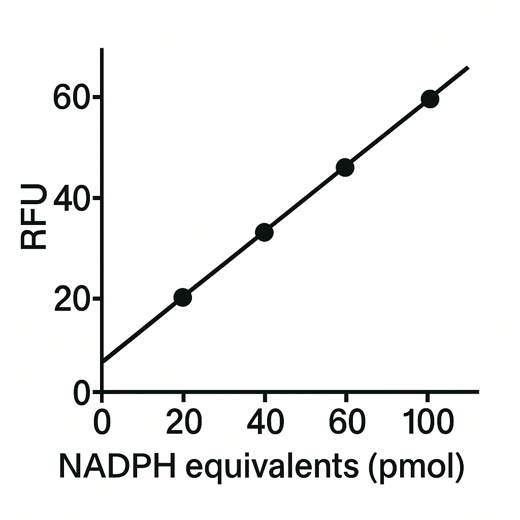
Prepare NADPH (or dye) standard curve in assay buffer (e.g., 0–2 µM range; adjust to your reader sensitivity).
-
In a black 96-well plate, add per well:
-
Assay buffer
-
Glucose (saturating or set for kinetic titrations)
-
ATP (saturating or controlled)
-
NADP+ (for dehydrogenase coupling)
-
Coupling enzyme(s)
-
Sample or control
-
-
Pre-incubate 5–10 min; then start reactions at 25–37 °C.
-
Kinetic read: measure fluorescence every 30–60 s for 10–30 min or end-point after a fixed time; ensure linear initial velocity.
-
Subtract blank (no enzyme) signals. Convert RFU to product units via standard curve.
-
Express activity as pmol/min/mg (or mU/mL).
Fluorescence measurement fundamentals and instrument training: University of Washington: https://www.washington.edu/; UC Berkeley: https://www.berkeley.edu/.
Kinetics and data analysis
-
Determine initial velocities from linear regions only.
-
For Michaelis–Menten parameters with respect to glucose (or ATP), fit v vs [S]; for GCK, consider sigmoidal behavior (Hill-type cooperative fit) if applicable.
-
For standard-curve mapping, use linear or 4PL depending on probe response. Curve-fit workflows and weighting strategies are detailed in Assay Guidance Manual (NIH Bookshelf): https://www.ncbi.nlm.nih.gov/books/NBK53196/.
-
Record instrument settings (gain, integration time, Ex/Em filters), and maintain audit trails per NLM data best practices: https://www.nlm.nih.gov/.
If you validate plate-reader linearity or optical stability, consult NIST SRMs and method notes for traceability: https://www.nist.gov/srm.
Controls and quality criteria
Include at minimum:
-
Blank (no enzyme; full reagents)
-
Zero-substrate control
-
Positive control enzyme or calibrated lysate
-
Matrix spike (spiked NADPH or fluorophore recovery)
Acceptance targets (typical research practice):
-
Intra-assay CV ≤ 10–15%, inter-assay CV ≤ 15–20%
-
Spike-recovery 80–120%
-
Dilution linearity/parallelism 80–120%
Plate layout, Z’-factor, and edge-effect mitigation strategies are summarized in the NIH Assay Guidance Manual: https://www.ncbi.nlm.nih.gov/books/NBK53196/.
Interference and troubleshooting
Interference sources
-
Autofluorescent compounds in lysates or media.
-
High phosphate, chelators, or detergents that inhibit GCK or coupling enzymes.
-
Air bubbles or particulates that scatter excitation light.
Troubleshooting map
-
Low signal → confirm substrate concentrations, enzyme integrity, and instrument gain; verify NADP+ and ATP stock quality (PubChem entries above).
-
High background → increase blank subtraction, check reagent contamination, and inspect plate for dust/particulates (university clean-lab tips via MIT OCW: https://ocw.mit.edu/).
-
Nonlinear kinetics → shorten read window to initial rate; ensure temperature stability (bench practices at Stanford: https://www.stanford.edu/).
-
Plate-to-plate drift → re-validate calibration with NIST-traceable references: https://www.nist.gov/programs-projects.
Reporting format (reproducibility checklist)
-
Reagent lots, buffer composition, and pH.
-
Plate type (black, low-bind), reader model, Ex/Em settings.
-
Standard curve source (NADPH or dye), range, and fit model.
-
Blank subtraction method.
-
Units and normalization basis (mg protein; µg lysate).
-
Raw data tables with time stamps, velocity calculations, and QC outcomes.
Data stewardship guidance: NIGMS reproducibility pages: https://www.nigms.nih.gov/, NLM information management: https://www.nlm.nih.gov/.
Safety & waste (general lab practice)
-
Wear appropriate PPE; follow local SOPs.
-
Manage dyes, peroxides, and organic solvents per institutional guidelines and OSHA Laboratory: https://www.osha.gov/laboratory.
-
Dispose of chemical waste per EPA resources: https://www.epa.gov/hwgenerators.
-
For biosafety workflows, see CDC laboratory practices: https://www.cdc.gov/lab/.
Learning resources (.edu/.gov quick list)
-
NIH/NCBI Gene: https://www.ncbi.nlm.nih.gov/gene/
-
NIH Bookshelf (Assay Guidance Manual): https://www.ncbi.nlm.nih.gov/books/NBK53196/
-
PubMed method searches: https://pubmed.ncbi.nlm.nih.gov/
-
NIST SRM & measurement science: https://www.nist.gov/srm and https://www.nist.gov/programs-projects
-
NLM/MeSH terminology: https://www.nlm.nih.gov/mesh/
-
University method hubs: MIT https://ocw.mit.edu/, Stanford https://www.stanford.edu/, UCSF https://www.ucsf.edu/, Harvard https://www.harvard.edu/, UW https://www.washington.edu/, UMich https://www.umich.edu/, UWisc https://www.wisc.edu/, UAz https://www.arizona.edu/, Berkeley https://www.berkeley.edu/
(These links expand on optics, enzymology labs, kinetics, and instrument best practices without clinical claims.)
SEO playbook for “Glucokinase Activity Assay Kit (Fluorometric)”
Primary keywords (use naturally in H1/H2 and alt text):
Glucokinase Activity Assay Kit (Fluorometric), glucokinase activity measurement, GCK enzyme kinetics, NADPH fluorescence, Ex/Em 340/460, fluorometric microplate assay, enzyme activity quantification, 96-well fluorometric assay, research use only, 4PL standard curve.
Title tag (≤ 60–65 chars):
Glucokinase Activity Assay Kit (Fluorometric) | Research-Grade
Meta description (≤ 155–160 chars):
Fluorometric glucokinase activity kit for research. Kinetic readouts, NADPH fluorescence, robust controls, and reproducible data analysis.
H1/H2 ideas:
-
H1: Glucokinase Activity Assay Kit (Fluorometric) — Research Use Only
-
H2: Principle, Coupling Chemistry, and Ex/Em Settings
-
H2: Kinetics, Standard Curves, and Data Quality
-
H2: Interference Controls and Troubleshooting
Image alt text templates:
-
alt="Glucokinase fluorometric assay standard curve (NADPH RFU)" -
alt="GCK enzyme kinetics initial velocity plot (v vs [glucose])" -
alt="96-well plate layout for glucokinase activity measurement"
Internal linking ideas:
-
Link to microplate readers, black plates, fluorescence standards, NADPH standards, G6PDH coupling reagents, and sample preparation buffers in your catalog.